[ad_1]
An international investigation coordinated by the Cancer Center Clinic University of Navarra (CCUN) has developed a diagnostic method that detects lung cancer early in blood samples. The study has identified a group of signals present in the cells of the immune system that improve the sensitivity of liquid biopsy in the early diagnosis of these patients.
These are the first conclusions of the AIR Projecta lung cancer screening study that has collected samples from more than 750 high-risk people at the University of Navarra Clinic and which have been analyzed in the Cima laboratories.
Liquid biopsy is a non-invasive technology capable of diagnosing different types of cancer through a blood test. However, the Clinic has reported, it still shows limitations in detecting the disease in its initial phases. “When a tumor develops, some cells of the immune system (lymphocytes) react to the cancer cells, multiplying and circulating through the blood. Until now, a single tumor recognition signal was sought in the lymphocyte. The novelty of this work is that we detect complete groups of signals (receptor repertoires), which are associated with the presence of cancer,” explains Dr. Luis Montuenga, director of the Lungsearch Group, dedicated to lung cancer, at the Cima University of Navarra and co-director of the study.
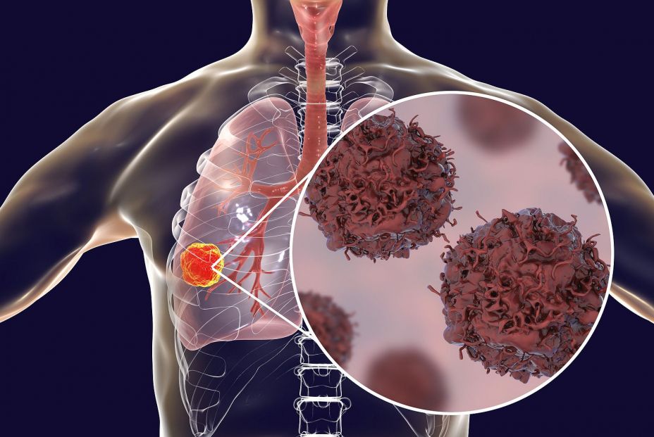
These repertoires of receptors can be identified through a molecular technique applied to blood analysis, which would open the door to very early detection of tumors and in a non-invasive way, the center has reported. Furthermore, combining this technique with other biomarkers, such as circulating DNA or specific proteins, would increase the sensitivity of this procedure. The results have been published in the scientific journal NPJ Precision Oncologyfrom the group Nature.
As indicated by Dr. Luis Seijo, director of the Department of Pulmonology at the Clínica Universidad de Navarra and specialist in the Lung Cancer Area of the CCUN, “these biomarkers can be useful in the context of screening, especially in three fundamental elements: in the characterization of the patient’s risk and those nodules that can be detected by CT, in the selection of patients and, potentially, even also in the prognosis.”
[ad_2]
Source link